
All of this, I'll tell you, begins with an attempt to waste time without really wasting it (trading science here for science there).I'm supposed to be studying for a genetics exam coming up this week. But when I look through the lecture presentation files, I keep getting distracted by the images in the slides. They're stunning: tiny little pieces of art that you (probably) never would have guessed were there.
One of the obvious problems of molecular genetics is that everything is so frustratingly small. To tackle this issue, scientists invent very creative ways to bring their subjects into view. Usually their methods rely on the type of elegant, futuristic technology that breezes over heads. But the people who turn away at this point are missing out.
They didn't necessarily need to know the technical, scientific details of the images to appreciate the intrinsic artistry that they represent (though it's pretty interesting to find out what you're looking at). Each bit of data doubles as an aesthetic micro-masterpiece, a collaborative effort of nature and human ingenuity. Every time we look at one of these images, it reminds us of the beauty and complexity of what exists beyond our normal perceptions. We're free to admire their forms but also, I think, to feel energized by our growing ability to identify the biological mechanisms behind the artwork.
This is the image that halted all my note-taking in class. Fluorescent proteins were used to identify synaptic circuits in a mouse's nervous system. The researchers cleverly coined their technique, "Brainbow". Their findings are important because they can use up to 90 unique color markers, and the method allows for tissue analysis without killing the organism. Even without knowing what these images say (and to a large extent, I don't either), you can admit that they're pretty brilliant to look at.

Dentate gyrus: Jackson Pollock or neurons?
The fruit fly, a favorite among geneticists, also has an attractive side. Gene expression, or how any gene translates the information encoded in its DNA into a useful end product, is a major topic of study in fruit fly embryo development. Scientists can inject dyes that will bind specifically to certain gene identifiers and not to others, which can create neat patterning effects in the resulting images. For example, 'pair-rule genes' are responsible for the segmentation of certain insects (like the fruit fly). Pair-rule genes divide the body along the anterior-posterior axis, promoting development of various structures like bristles to form in alternating segments along the body. The images show interesting dye stripe patterns where each pair-rule gene is being expressed.

Anterior-Posterior Patterning in Fruit Fly Embryo
Fluorescent markers, like those used in the 'Brainbow', can also be helpful. The photo below shows the location of different proteins in heart and muscle cells throughout a fruit fly embryo.

From the National Heart, Lung, and Blood Institute (NIH)

Both are pretty far cries from how we typically
envision the fruit fly.
Sometimes, though, these photographs are created with no exact scientific end in mind. For instance, powerful electron microscopes can make everyday objects look ethereal and extraordinary.
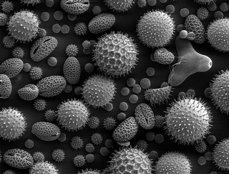
Pollen.

Snowflakes.

Doxorubin in methanol and dimethylbenzenesulfonic acid (80x)
Lars Bech, 1996

Cross-section of a very young beech (40x)
Jean Rüegger-Deschenaux, 1994

Double transgenic mouse embryo, 18.5 days (17x)
Gloria Kwon, 2007

Deformation of a polyethylene folio (40x)
Zdenka Jenikova, 2002

Anopheles (mosquito) eye (20x)
Marie Andersson, 2010

Made by John Hart, using a scanning electron microscope
and millions of carbon nanotubes produced
in a chemical reaction.

Hmm...
The opposite of art is science? Well, I don't know about that.
P.S. This is my favorite site for science as art.
Song of the Day
Why? (Live)
Andrew Bird*
*If you're like me and you just can't get enough biology allusions and/or Andrew Bird, check out 'Imitosis'.
No comments:
Post a Comment